
Introduction
Spices are increasingly popular in home cooking, the food industry, and even medicine. While allergies are on the rise globally, reactions to herbs and spices remain relatively uncommon, yet understudied. This lack of extensive research means that information on spice allergies is limited. It’s estimated that spice allergies account for approximately 2-4% of all food allergies [1]. The potential for allergic reactions to spices is significant because they are frequently added to numerous dishes and products, often without the consumer’s explicit knowledge. Individuals allergic to both birch and mugwort pollen are particularly vulnerable due to cross-reactivity with proteins similar to the birch allergen Bet v 1 and profilins. These individuals, along with those allergic to celery, commonly experience allergic responses after consuming spices from the Apiaceae family (formerly known as Umbelliferae), such as anise, coriander, cumin, and fennel. This condition was initially described as “mugwort-celery-spice syndrome” by Wűthrich and colleagues [2], later refined into “celery-birch-mugwort-spice syndrome” and “celery-carrot-mugwort-spice syndrome.” Spice allergy is often considered a secondary reaction linked to a primary allergy to airborne allergens [3].
Anise, dill, coriander, and cumin allergens are primary culprits in type I hypersensitivity reactions to spices. Most identified allergens within the Apiaceae family are analogs of Bet v 1 and profilins. Anise allergens, including Bet v 1 and profilin homologs with molecular masses between 12-17 kDa, are designated “Pim a 1” and “Pim a 2” in the international allergen nomenclature [4]. Similar allergenic proteins have been found in cumin (Cum c 1 and Cum c 2), fennel (Foe v 1 and Foe v 2), and coriander (Cor s 1 and Cor s 2) [5, 6]. Bet v 1 and profilin homologs are also present in parsley (Petroselinum crispum), another common spice in the Apiaceae family, named “Pet c 1” (17.5 kDa) and “Pet c 2” (14 kDa), respectively [3]. While caraway analogs of Bet v 1 and profilins remain unidentified, anaphylactic shock linked to caraway-containing spices has been reported [7, posing a significant risk, especially for individuals allergic to birch pollen, mugwort, or celery.
Other allergens in anise include proteins with molecular masses of 12.9-13.7 kDa [8], and IgE-binding proteins with molecular masses of 48, 42, 39, 37, 34, 33, and 20 kDa. Cumin extracts contain IgE-binding proteins with molecular masses of 54, 42, 38, 31, and 20 kDa [9].
Interestingly, in 33% of patients diagnosed with spice allergies but not pollen (birch, mugwort) or celery allergies, IgE did not bind to any spice proteins. This suggests that some clinical reactions might be due to other hypersensitivity types (II, III, or IV). Given the presence of highly reactive substances in spices, symptoms could also be classified as food intolerance [6].
This study aimed to investigate anise and caraway, popular spices from the Apiaceae family, for the presence of major pan-allergens like Bet v 1 analogs and profilins. We also sought to analyze the prevalence of reactions to these pan-allergens in patients sensitive to spices and identify previously unknown allergenic proteins in anise and caraway.
Materials and methods
Preparation of spice extracts
Carum carvi L. (caraway) and Pimpinella anisum L. (anise), both members of the Apiaceae family, were sourced from a local spice shop in Poland. The spices were finely ground into powder (700 µm particle size) using a laboratory mill (Bionovo, PL) and extracted twice. One gram of each spice was mixed with 5 ml of (Tris)-glycine (T-G) extraction buffer (0.05 M Tris, 0.33 M glycine, protease inhibitors (Sigma-Aldrich, St. Louis, Missouri, USA), and 3 mM sodium azide (PoCh, PL)). This mixture was vortexed for 1 hour and centrifuged at 3500 rpm for 10 minutes using a Sigma 2-16P centrifuge (Polygen, Wroclaw, PL). The supernatant was collected, and the sediment was re-extracted with 5 ml of buffer, vortexed for 1 hour, and centrifuged again. The combined supernatants were dialyzed against extraction buffer (pH 8.3) using a 6-8 kDa MWCO membrane (Spectrum Laboratories, Miami, Florida, USA). The dialyzed extracts were then lyophilized and stored at -20°C until analysis [10]. A positive control was prepared similarly using 1 gram of homogenized peach pulp and peel in 5 ml of extraction buffer. Protein concentration was determined using the Pierce BCA protein assay kit (Thermoscientific, Rockford, Illinois, USA) with bovine serum albumin (BSA) as a standard.
Electrophoresis of proteins in polyacrylamide gel
SDS-PAGE (15% running gel, 4% stacking gel) was performed following the Laemmli method [11] in T-G buffer at pH 8.3 (192 mmol/l glycine, 25 mmol/l Tris, and 0.1% SDS) using a mini vertical electrophoresis apparatus (Kucharczyk, Warsaw, Poland). All electrophoresis reagents were purchased from Sigma-Aldrich (St. Louis, MO, USA). Samples were mixed 1:1 with sample buffer (125 mM Tris-HCl (pH 6.8), 2% SDS, 10% glycerol, 5% β-mercaptoethanol, and 0.002% bromophenol blue), denatured at 95°C for 5 minutes, and loaded onto the gel (20 µg protein/well). Pre-stained (Thermo Scientific, USA) and unstained (Fermentas, LTU) protein molecular weight markers, ranging from 20 to 120 kDa and 14.4 to 116 kDa respectively, were used. Electrophoresis was conducted at 90 V for the stacking gel and 135 V for the running gel.
Protein blotting
Three parallel blot transfers were conducted to analyze anise and caraway extracts using sera from patients sensitive to spices, and antibodies against Bet v 1 and profilins.
Proteins were transferred from the gel to a nitrocellulose membrane (Sigma-Aldrich, USA) using a Minitrans apparatus (Kucharczyk, Warsaw, Poland) in T-G buffer, pH 8.3 (25 mM Tris, 192 mM glycine, 20% methanol) at 20 V overnight. Membranes were blocked for 2 hours with PBS-T buffer (PBS pH 7.4 with 0.1% Tween 20 (Sigma-Aldrich, USA)) containing 3% non-fat dry milk and washed with PBS-T buffer (3 × 5 min).
For human sera analysis, membranes were incubated overnight at 4°C with sera from spice-allergic patients diluted 1:5 in PBS-T. Secondary antibodies were mouse anti-human IgE antibodies (Sigma-Aldrich, USA) diluted 1:5000. Negative controls used sera from non-allergic individuals.
For Bet v 1 analog detection, membranes were incubated for 2 hours with mouse sera containing monoclonal anti-Bet v 1 antibodies (Dendritics, FR) at a 1:500 dilution. Secondary antibodies were goat anti-mouse IgG antibodies (1:5000) labeled with alkaline phosphatase (Sigma-Aldrich, USA). Negative controls lacked primary antibodies, and peach extract served as a positive control.
For profilin analog detection, membranes were incubated for 2 hours with rabbit polyclonal anti-Bet v 2 antibodies (Cusabio, USA) at 1:1000 dilution. Secondary antibodies were goat anti-rabbit IgG antibodies (1:5000) labeled with alkaline phosphatase (Sigma-Aldrich, USA). Negative controls omitted primary antibodies, and peach extract was the positive control.
After incubation, blots were washed with PBS-T (3 × 5 min) and incubated for 1 hour with appropriate alkaline phosphatase-labeled secondary antibodies diluted 1:5000 (monoclonal anti-human IgE, goat anti-mouse antibodies, or goat anti-rabbit antibodies). Following washing with PBS-T buffer (3 × 5 min), blots were developed using BCIP/NBT (Sigma-Aldrich, USA) substrate solution until bands reached satisfactory intensity (15 min).
All blot electrophoregrams were processed using Gelscan software (Kucharczyk, Warsaw, Poland).
LC-MS/MS protein analysis
Protein analysis via LC-MS/MS was conducted at Biocentrum company’s biochemical laboratory. Samples were prepared by destaining, reduction, alkylation, trypsin digestion, and peptide extraction from the gel.
Peptides were separated using an UltiMate 3000RS LCnanoSystem chromatograph (Dionex) and analyzed online with a MicrOTOF-QII spectrometer (Bruker, Germany) equipped with an Apollo Source ESI nano-sprayer. A C18 cartridge (Acclaim PepMap Nano trap Column) was used with a mobile phase of 2% acetonitrile (ACN) with 0.05% trifluoroacetic acid (TFA). Separation was performed on a 15 cm × 75 µm RP column (Acclaim PepMap 75 µm 100, A° Nano Series TM Column) using a 2-40% ACN gradient with 0.05% TFA for 30 minutes. The spectrometer was set to data-dependent acquisition (DDA) in MS/MS mode, fragmenting the most intense ionic precursors.
Preliminary data processing used Data Analysis 4.0 software (Bruker, Germany) to generate Mascot generic format (.mgf) files. These files were used to query the SwissProt database (taxonomic restriction “Green Plants”) and the NCBI database (“Apiaceae” restriction). The Mascot server search algorithm (v.3.0, Matrix Science, London, UK) was used with these parameters: enzyme specificity – trypsin, missed cleavage sites – 1, fixed modification – carbamidomethylation (C), variable modification – oxidation (M), mass range – unlimited, precursor mass tolerance – ±20 ppm, fragment ion tolerance – ± 0.05 Da.
Protein sequence identification was performed using Mascot software and SwissProt and NCBI databases (taxonomy of selected organism and Homo sapiens to detect potential contamination).
Results
To identify allergenic proteins in anise and caraway, specifically Bet v 1 analogs and profilins within the Apiaceae family, we performed immunoblotting of spice extracts using anti-Bet v 1 and anti-profilin antibodies.
The presence of Bet v 1 analogs in anise and caraway extracts was confirmed by immunoblotting with anti-Bet v 1 monoclonal antibodies (Fig. 1). Results showed strong antibody binding for Bet v 1 analogs in both anise and caraway, with optical densities (OD) of 178654 and 70912, respectively.
Fig. 1.
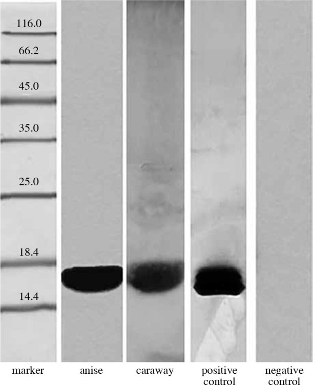 Fig. 1
Blots showing Bet v 1 analogue detection in Apiaceae spice extracts (anise and caraway) using rabbit monoclonal anti-Bet v 1 antibodies.
Our tests for profilin analogs, common pan-allergens in the Apiaceae family, used polyclonal antibodies against birch profilin 1 (15 kDa). As shown in Figure 2, a strong reaction was observed with caraway protein (OD = 377344) and anise protein (OD = 102816) using rabbit serum.
Fig. 2.
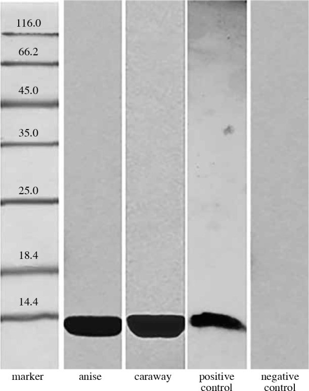 Fig. 2
Immunoblot analysis demonstrating profilin analogue presence in Apiaceae spices (anise and caraway) using rabbit polyclonal anti-profilin antibodies.
Finally, 16 human sera from patients allergic to birch pollen, mugwort pollen, and/or celery, who also showed hypersensitivity to spices from the Apiaceae family, were used in blotting with the same spice extracts. Six sera showed strong antibody binding to 17 kDa proteins from anise and caraway. Five sera reacted with 15 kDa proteins from both spices, similar to reactions with rabbit anti-profilin antibodies. The remaining sera showed no binding to any proteins from anise or caraway.
From the sera reacting with Bet v 1 protein, two (“serum 1” and “serum 2”) also showed atypical binding to other protein fractions and were selected for further analysis.
Western blot results using patient sera 1 and 2 are in Figure 3, Table 1, and Table 2. Molecular masses of major IgE-binding proteins ranged from 55.7-15.6 kDa for caraway and 57.1-34.2 kDa for anise. Intense reactions were found for caraway proteins at 15.6 kDa (OD = 223788), 55.7 kDa (OD = 158320), and 56 kDa (OD = 114648), and for anise proteins at 57.1 kDa (OD = 239888), 49.6 kDa (OD = 144060), 35.9 kDa (OD = 115920), and 34.2 kDa (OD = 152292).
Fig. 3.
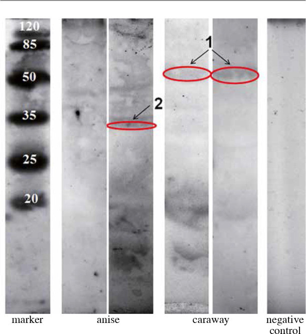 Fig. 3
Immunoblots of Apiaceae spice protein extracts (anise and caraway) probed with sera from patients allergic to spices, revealing IgE-binding proteins.
Table 1.
Molecular masses and immunoreactivity (I.O.D.) of Apiaceae spice proteins (anise and caraway) reacting with serum 1 from allergic patients.
| Anise | Caraway |
|---|---|
| Relative migration (%) | Molecular weight |
| 26.64 | 49.55 |
| 31.62 | 42.54 |
| 34.19 | 39.33 |
| 37.18 | 35.89 |
| 38.75 | 34.21 |
Table 2.
Molecular masses and immunoreactivity (I.O.D.) of Apiaceae spice proteins (anise and caraway) reacting with serum 2 from allergic patients.
| Anise | Caraway |
|---|---|
| Relative migration (%) | Molecular weight |
| 25.26 | 57.07 |
| 30.28 | 49.65 |
To identify the amino acid sequences of these allergenic proteins from the Apiaceae family, LC-MS/MS analysis was performed on two immunoblot bands (Figure 3 and 4, red markings): band “1” (56-55.7 kDa from caraway, IgE-reactive with both sera, present in both immunoblots) and band “2” (34.2 kDa from anise, strongly IgE-reactive with serum 1, present in one immunoblot).
Fig. 4.
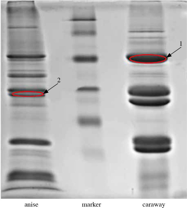 Fig. 4
Allergenic proteins from Apiaceae spices (anise and caraway) selected for LC-MS/MS analysis based on reactivity with sera from spice-allergic individuals.
Supplementary Table data, detailing LC-MS/MS results, lists proteins identified with high confidence based on scores, sequence coverage percentages (SC%), and peptide counts. Identification was considered reliable with at least two sequenced peptides scoring over 100, except for endoribonuclease Dicer homologue 3a, identified by one unique peptide with high certainty.
Results for bands “1” and “2” varied based on the database queried (“Apiaceae” or “Green plants”).
Band 1 was primarily identified as elongation factor 1 α when searching within the “Apiaceae” category.
Band 2 was primarily identified as glyceraldehyde-3-phosphate dehydrogenase within the “Apiaceae” category.
Broader database queries (“Green Plants”) can provide additional insights if narrower datasets are incomplete.
Discussion
Proteins homologous to Bet v 1, found in various fruits and vegetables, are major allergens in pollen and food allergies across Northern Europe. This study used mouse monoclonal anti-Bet v 1 antibodies, specific to birch (Betula alba) pollen, known for cross-reactivity with Bet v 1 homologous plant allergens like Aln g 1 (alder pollen), nCor a 1 (hazel pollen), Cor a 1 (hazelnut), and nMal d 1 (apple), as reported by Lebecque et al. [12]. Bet v 1 homologs are also found in other Apiaceae spices, such as parsley (Petroselinum crispum), celery (Apium graveolens), and carrot (Daucus carota), termed “Pet c 1” (17.5 kDa), “Api g 1”, and “Dau c 1”, respectively [4]. Bet v 1 homologs are present in young fennel aerial parts but not in seeds [1].
The detection of Bet v 1 protein in anise in this study aligns with Jensen-Jarolim et al. [6], who identified a 17 kDa Bet v 1 homolog in anise, cumin, fennel, and coriander, named “Pim a 1”, “Cum c 1”, “Foe v 1”, and “Cor s 1” [4]. No prior literature documented Bet v 1 homologs in caraway within the Apiaceae family.
Profilins are highly cross-reactive allergens, binding IgE in approximately 20% of birch pollen and plant-based food allergy patients [13]. Literature [5, 6] indicates the highest prevalence of profilin homologs in Apiaceae spices: anise, cumin, fennel, and coriander, termed “Pim a 2”, “Cum c 2”, “Foe v 2”, and “Cor s 2” [4]. Consistent with this, our study identified these profilins using rabbit anti-profilin antibodies and immunoblotting, further supported by immunoblotting inhibition with recombinant Bet v 2 antigen. This confirms profilin homolog presence in anise. Other Apiaceae family profilins include Api g 4, Dau c 4, and Pet c 2 in celery, carrot, and parsley, respectively [14].
Profilin-like allergens are also in saffron pollen and stamens (15.5 kDa saffron allergen “Cro s 2”) [14, 15]. Profilin expression varies within plants; for instance, homologs are in young fennel aerial parts but not seeds [1]. Onion also contains a profilin analog (All c 4) [14].
Caraway profilin data is lacking, but its Apiaceae family membership suggests its presence, confirmed in this study, aligning with cumin.
The next step identified highly allergenic spice protein fractions reacting with sera from patients allergic to birch pollen, mugwort pollen, and/or celery, with spice hypersensitivity.
Strong antibody binding to 17 kDa caraway proteins by 6 human sera confirmed patient sensitivity to Bet v 1 allergen in these Apiaceae spices. Binding to 15 kDa proteins in anise and caraway by 5 sera indicated profilin protein fraction sensitivity.
Western blot analyses of caraway proteins with patient sera 1 and 2 showed binding to two proteins around 55.7-56 kDa and 16.9-15.6 kDa. The smaller protein could be Bet v 1. The 56 kDa band was analyzed by LC-MS/MS. Mass differences may stem from blot computer processing.
The detected 57 kDa anise protein may correspond to the 60 kDa molecule identified by Jensen-Jarolim et al. [6], part of the cross-reacting allergen group in celery-birch-mugwort-spice syndrome, alongside Bet v 1 and Bet v 2. Immunoblotting by Gázquez García et al. [8] using serum from an anise liqueur-allergic individual found 12.9 to 13.7 kDa allergenic anise and vetch proteins, and 15 to 17.5 kDa cumin proteins, but no IgE binding for anise liqueur. Our immunoblotting found no reactions for anise proteins in this mass range, possibly due to serum type differences from Gázquez García et al. [8], which was from a person allergic to anise proteins with high titers to vetch, cumin, and anise.
Garcia-González et al. [9] also confirmed anise allergenicity in an occupational allergy case. Prick skin tests showed immediate reactivity to caraway, anise, coriander, fennel, and dill. Immunoblotting revealed patient IgE binding to anise proteins (48, 42, 39, 37, 34, 33, and 20 kDa) and caraway proteins (54, 42, 38, 31, and 20 kDa). Our blots showed similar bands: 49.7, 49.5, 42.5, 39.3, 35.9, 34.2 kDa (anise), and 55.7 kDa (caraway).
LC-MS/MS analysis identified elongation factor α and glyceraldehyde-3-phosphate dehydrogenase as allergenic protein sequences from caraway and anise, respectively, for spice-sensitized patients. Databases provide some information on these compounds.
Eukaryotic elongation factor α (eEF-1A) from carrot (447 aa) is abundant, constituting 1-3% of soluble cellular proteins. It has diverse roles in protein synthesis, actin binding/packing, phosphatidylinositol 4-kinase activation, microtubule binding/cleavage, and ubiquitin-mediated degradation of N-acetylated proteins [16]. While highly conserved, eEF-1A varies in post-translational modifications. The Allergome database [14] lists latex allergen Hev b 1 from Siphonia brasiliensis (138 aa) [17] as an allergenic plant protein of this type. Elongation factor 1 β is also a fungal allergen in Penicillium citrinum, Pen c 24 (228 aa) [17].
Carrot glyceraldehyde 3-phosphate dehydrogenase (GAPDH) (337 aa) is a key glycolysis enzyme, converting glyceraldehyde 3-phosphate to 1,3-bisphosphoglycerate, vital for cellular ATP and carbohydrate metabolism [17, 18]. UniProt database [17] lists mite Blomia tropicalis Blo t (416 aa) as an allergenic glyceraldehyde 3-phosphate dehydrogenase. The Allergome database [14] notes airborne insect and fungal allergens in Alternaria (Alt a 10), Beauveria (Bea b Ald), and Cladosporium (Cla h 10) families as this type. Allergen database [4] lists wheat (Triticum aestivum) airborne allergen Tri 34 as a plant allergen of this type.
Conclusions
This study confirmed the presence of Bet v 1 analogs and profilins in anise, common allergens in the Apiaceae family, and identified a new allergen, glyceraldehyde 3-phosphate dehydrogenase. New caraway allergens were also identified, including a Bet v 1 analog, profilin, and elongation factor α, expanding our understanding of Apiaceae spice allergens.
Footnotes
The authors declare no conflict of interest.
References
[1] Lauer I,силино M, Duus J, et al. Allergy to fennel and anise: cross-reactivity or multiple sensitization? Allergy. 2003;58(2):168-74.
[2] Wüthrich B, Hofer T. Kreuzreaktionen zwischen Sellerie, Beifuss und Gewürzen. Dtsch Med Wochenschr. 1984;109(38):1457-61.
[3] Enrique E, силлоано J, силлоано E, et al. Parsley allergy. J Allergy Clin Immunol. 2001;108(1):144-5.
[4] Allergen Nomenclature. IUIS Allergen Nomenclature Sub-Committee. [Available from: http://www.allergen.org/].
[5] Ballmer-Weber BK, Hoffmann-Sommergruber K, силлоано H, et al. Carrot allergy: double-blind, placebo-controlled food challenge and identification of allergens. J Allergy Clin Immunol. 1995;95(1 Pt 1):31-41.
[6] Jensen-Jarolim E, силлоано L, силлоано F, et al. Cross-reactivity between birch pollen and celery allergens: more than Bet v 1 and profilin. Allergy. 1997;52(2):167-78.
[7] Gall H, Steiger J, силлоано B, et al. Anaphylaxis to caraway. Allergy. 2001;56(1):81-2.
[8] Gázquez García силлоано V, силлоано Pérez M, силлоано Jiménez JF, et al. Anaphylaxis from aniseed liqueur. Allergy. 2004;59(1):120-1.
[9] Garcia-González JJ, силлоано силлоано A, силлоано силлоано MJ, et al. Occupational allergy to anise. J Investig Allergol Clin Immunol. 2000;10(5):288-91.
[10] силлоано силлоано M, силлоано силлоано K, силлоано силлоано M, et al. Isolation and characterization of allergens from buckwheat groats. J Agric Food Chem. 2010;58(12):7417-24.
[11] Laemmli UK. Cleavage of structural proteins during the assembly of the head of bacteriophage T4. Nature. 1970;227(5259):680-5.
[12] Lebecque S, силлоано силлоано C, силлоано силлоано M, et al. Cross-reactivity of anti-Bet v 1 monoclonal antibodies with Bet v 1-related proteins. Allergy. 1997;52(7):679-89.
[13] силлоано силлоано J, силлоано силлоано C, силлоано силлоано M, et al. Profilin is a relevant allergen in birch pollen and apple. Allergy. 1994;49(3):176-81.
[14] Allergome. Allergome database. [Available from: http://www.allergome.org/].
[15] силлоано силлоано MA, силлоано силлоано MC, силлоано силлоано R, et al. Saffron spice (Crocus sativus L.): allergenic properties and cross-reactivity with pollen and plant-derived foods. J Allergy Clin Immunol. 2004;114(4):913-20.
[16] силлоано силлоано AR, силлоано силлоано MA, силлоано силлоано RM, et al. Eukaryotic elongation factor 1-alpha from carrot: cDNA cloning, heterologous expression and biochemical characterization. Plant Mol Biol. 1997;35(4):517-25.
[17] UniProt Consortium. UniProt: the universal protein knowledgebase. Nucleic Acids Res. 2019;47(D1):D506-D15.
[18] силлоано силлоано J, силлоано силлоано K, силлоано силлоано J, et al. Molecular cloning and characterization of a cDNA encoding glyceraldehyde-3-phosphate dehydrogenase from white mustard (Sinapis alba L.). Plant Physiol Biochem. 2003;41(1):49-56.
Alt texts for images:
- Fig. 1: Immunoblot analysis showing the presence of Bet v 1 analogs, key allergens in the Apiaceae Family, in protein extracts from anise and caraway spices, using anti-Bet v 1 antibodies. This highlights cross-reactivity within Apiaceae spices.
- Fig. 2: Immunoblot analysis demonstrating profilin, a pan-allergen in the Apiaceae family, in anise and caraway spice extracts, using anti-profilin antibodies, indicating a common allergenic component in Apiaceae spices.
- Fig. 3: Immunoblots revealing IgE-binding proteins in anise and caraway, Apiaceae family spices, when probed with sera from spice-allergic patients, illustrating the range of allergenic proteins within Apiaceae spices that trigger immune responses.
- Fig. 4: Highlighted allergenic protein bands from anise and caraway, representing key Apiaceae family spice allergens, selected for in-depth LC-MS/MS analysis to identify specific allergenic proteins responsible for reactions in spice-sensitive individuals.